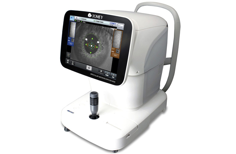
Our Equipment

Eye Clinic for Animals

ECA operates out of a brand new state of the art Veterinary Hospital dedicated to ophthalmology located in Artarmon.

ECA also operates out of the RSPCA Veterinary Hospital in Yagoona.

Our goal is to have the most advanced equipment available in veterinary and also human ophthalmology.

As a result of being world leaders in corneal transplantation, ECA has purchased equipment that no other private practice in Australia, and possibly the world have.

This high-end equipment is used extensively in human ophthalmology. In our desire to be at the absolute cutting edge of all things ophthalmology, ECA has made a significant capital outlay to stay at the diagnostic and therapeutic forefront.

OUR EQUIPMENT
treatment image

Tomey EM 4000 Specular microscope. This machine allows us to assess the viability and number of endothelial cells on the back of the cornea. This is especially useful in corneal transplant surgery to assess the viability of prospective donors, as well as monitor the pre and post operative condition of recipient patients. This machine is also very useful prior to cataract surgery in patients over 8-10 years old. In this way ECA can optimize the surgical outcomes after cataract removal.

treatment image

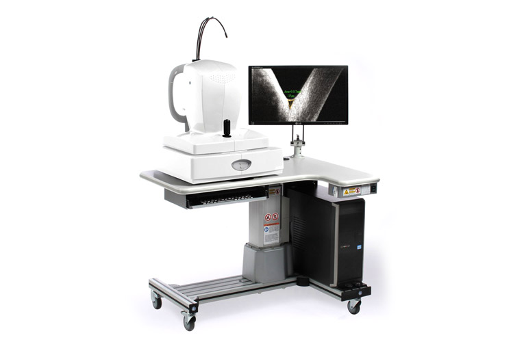
Optovue - Rtvue Optical Coherence Tomographer (OCT). This machine is a non-invasive imaging test and scans the eye without touching it. It uses light waves to take cross section images of the cornea, lens and retina. In ECA we use it especially to accurately assess the corneal width, postoperative location of transplanted corneal endothelium, and for assessment of the retina in cases of vision loss or deterioration.

treatment image

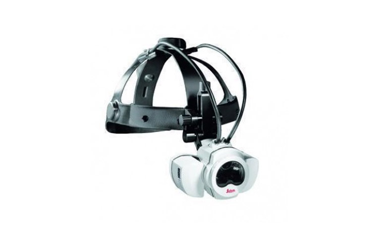
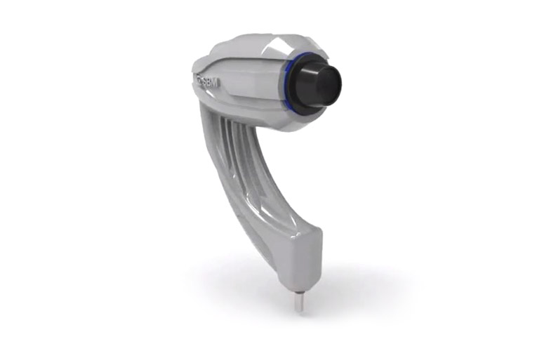
Infrared 810nm laser (Iridex Oculight SL) The specific wavelength of light of 810nm is selectively absorbed by pigmented tissue. Therefore, this laser can be utilised to selectively destroy pigmented tissue within the eye. This is helpful to treat a whole raft of conditions, including iridial cysts or iridal melanomas, or destroying the portion of the eye responsible for the generation of aqueous humour to prevent glaucoma. The headset attachment (shown below), offers us the ability to do retinal surgery without necessarily entering the eye in some cases.

treatment image

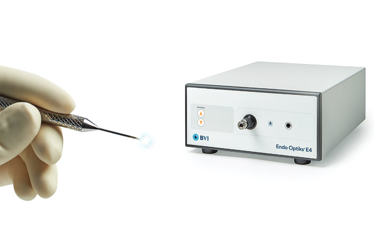
Endolaser (Iridex Oculight SL and BVI Endo Optiks E4 Ophthalmic Endoscopy System). Glaucoma can be treated surgically via the application of energy to destroy the ciliary body epithelium, responsible for the production of aqueous humour. There are several ways to do this, however, in humans the use of an endoscopic camera with a laser-attached to visualise and manually destroy the ciliary body epithelium is the modality of choice. This allows for wide field-of view, maneuverability, and real-time footage within the eye during the time of surgery. We too have the capability to perform this procedure, which has many theoretical benefits to other available techniques that are possible without this endoscopy unit.

OPERATING MICROSCOPES

We have a fleet of operating microscopes (6 in total), which allow us to perform the finest of ocular surgeries under magnification. As the nature of ophthalmic surgery is very delicate, using fine structures, we at ECA have strived to ensure that we have the best surgical facilities possible.

treatment image

“Lucy” - The Lumera 700 operating microscope with vitreoretinal upgrade The arrival of “Lucy” in June 2020 is the jewel in the ECA crown. The Lumera 700 is one of the world’s best operating microscope units and is used extensively for high end human ophthalmic surgery. This microscope allows for complete surgical capabilities of the entire eye, including vitreoretinal surgery with motorised vitreoretinal capabilities.

treatment image

“Victor” - The Visu 200 operating microscope. The Visu 200 has been used extensively in human ophthalmology hospitals and has proven to be a reliable workhorse for ECA. With the functional ability to do everything that we require in ECA, including complex eyelid surgeries, corneal transplants, cataract surgeries and retinal surgeries, Victor has been the backbone of ECA surgical practice for the last few years.

treatment image

Scan Optics - 5000 SE Portable Operating Microscope with Coaxial Video System. This Australian made portable operating microscope offers great versatility for all of our surgeries we do outside of the main clinics. This includes work at Taronga Park Zoo, Western Plains zoo in Dubbo, and all our horse surgery at various locations throughout Sydney. A nice addition especially when we need exacting depth of field and focussing control such as in horse cataract surgery.

treatment image

Leica HM500 Headmounted Surgical Microscope This is the world's only head mounted surgical microscope. It combines the flexible, mobile properties of magnifying glasses with the magnification capabilities of surgical microscopes. It is an amazing piece of equipment that can run off a battery but still retains the capacity for automatic focus and foot operated magnification. Fantastic in places where we can’t easily get electricity in the field, such as Rhinoceros surgery in a paddock at Western Plains Zoo.

Ultrasound Pachymetry

Ultrasound pachymetry is used to measure the corneal thickness, and traditionally is used as an indirect screening option to assess corneal endothelial cell function. The benefit over more sophisticated testing of the cornea such as Tomey EM 4000 specular microscopy or Optovue-Rtvue ocular coherence tomography is the portability of the handheld pachymeters.

ECA has two types of portable pachymeters.

treatment image

The Accutome Pachpen is ideal to assess corneal thickness in our outpatient population. With market leading lightness and ergonomic design it is an easy to use screening tool. This machine allows us to assess corneal thickness from 300 microns to about 1000 microns.

treatment image

The DGH-555B Pachette 4 from DGH technologies is an especially useful piece of equipment that has been specially modified for our veterinary use. Bigger than the handheld Accutome Pachpen, it is table mounted but still very user friendly. This machine allows us to assess corneal thickness from as thin as 100 microns (1/10 th of a millimeter !!) to 2000 microns (2 mm), important in the pre and postoperative monitoring of our corneal transplant patients.

treatment image

BX53F2 Microscope for transmitted and reflected light with a high definition DP 74 20 Mega Pixel Camera and a 24 inch ultrawide screen, with industry leading cellSens software for digital image acquisition and manipulation. This microscope is fundamental to our assessment of the number and viability of endothelial cells in the donor tissues retrieved. With brilliant optical clarity and precision image acquisition, this is an essential tool for our corneal endothelial transplant work. Furthermore the cellSens software package allows manual and fully automatic image acquisition and date analysis.

treatment image

Ocular Ultrasound (Accutome B-Scan Plus) A B-wave ultrasound of the eye allows for an assessment of the gross structures of the eye, and assess the dimensions of the gross structures of the eye. We have the benefit of high quality B-scan ultrasound probes to allow us to assess the internal ocular structures.

treatment image

Ultrasound biomicroscopy (Accutome UBM Plus) An ultrasound biomicroscope generates a high frequency, low penetrance ultrasound wave that allows for in vivo imaging of ocular tissues. The high resolution of this technology allows for visualisation of ocular structures with clarity that previously was only possible in tissues that have been surgically removed. This allows for the assessment of the cornea, anterior chamber, iridocorneal angle and anterior iris.

treatment image

Ocular Surface Analysis (ICP VET SBM Sistemi) Tears have many important roles in establishing corneal health. Crudely, the tear film can be divided in to three components, the lipid layer, the aqueous component and a thick mucous layer. All three layers are crucially important in maintaining corneal health, and allowing for the health of the cornea. This diagnostic suite allows for the assessment of the individual components of the tear film, and assess the tear film’s components.

treatment image

RETevet™ Portable ERG The RETevet™ portable ERG offers an intuitive interface that allows veterinary ophthalmologists and researchers to perform electroretinograms (ERGs) to measure the retinal function of animals.

OUR LOCATIONS

OUR OPENING HOURS: 9am – 5pm

ARTARMON

ARTARMON
53-55 Herbert Street, Artarmon NSW 2064

Ph: (02) 9058 3500
9am - 5pm

info@eca-aus.com.au

YAGOONA

YAGOONA
201 Rookwood Road Yagoona NSW 2199 (Inside RSPCA Veterinary Hospital)

Ph: (02) 9058 3500
9am - 5pm

info@eca-aus.com.au

BOOK AN APPOINTMENT
Online Booking