
What is dry eye disease?
Dry Eye Disease, also known as Keratoconjunctivitis Sicca (KCS), was once thought to be caused simply by a lack of tear production to lubricate the cornea (the clear front part of the eye). However, we now understand that tears are a complex fluid made up of different components, each playing a crucial role in maintaining corneal health and preventing infections from bacteria and fungi.
Because of this, Dry Eye Disease is now classified into two types:
- Quantitative Dry Eye – When the eye doesn’t produce enough tears.
- Qualitative Dry Eye – When the tear composition is abnormal, affecting the quality and function of the tears.
Both forms can lead to irritation, inflammation, and discomfort, requiring proper diagnosis and treatment to protect long-term eye health.
What Makes Up the Tear Film?
The tear film (or tear broth) is made up of three essential layers, each playing a crucial role in protecting and maintaining the health of the eye:
- Lipid Layer – This oily layer is produced by the Meibomian glands in the eyelids. Its main function is to slow down tear evaporation, helping to keep the eye hydrated for longer.
- Aqueous Layer – The watery component of the tear film, produced by the lacrimal glands in the upper eyelid and nictitans (third eyelid). This is the largest part of the tear film and helps flush away debrisand microorganisms, keeping the cornea clean and protected.

Each of these layers works together to ensure proper eye lubrication, clear vision, and protection frominfections. Any disruption in quantity or quality can lead to Dry Eye Disease (KCS) and discomfort for the affected dog.
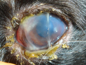
How Dry Eye Affects the Eye
As mentioned earlier, Dry Eye Disease (KCS) can be caused by either low tear production (Quantitative Dry Eye) or poor tear quality (Qualitative Dry Eye). While this classification helps in diagnosing the condition, it’s also important to understand how the lack of functional tears impacts the eye over time. Without a healthy tear film, the cornea struggles to protect and nourish itself.
To compensate, the eye undergoes several changes:
- Thickening of the cornea as it tries to reinforce itself
- Growth of blood vessels across the surface in an attempt to deliver more nutrients
- Pigment buildup that further affects vision
Although these changes help the eye survive, they come at a cost. The cornea becomes less transparent, vision becomes impaired, and the eye remains in a state of chronic discomfort. This is why early diagnosis and proper treatment are essential to managing the disease and preventing long-term damage.
What Causes the Two Types of Dry Eye Disease?
Quantitative Dry Eye Disease (lack of aqueous tear production) is typically caused by one of three underlying issues:
- Congenital Lacrimal Gland Hypoplasia or Aplasia – Some dogs are born with underdeveloped or absent lacrimal glands, meaning they produce little to no tears from birth.
- Autoimmune Dry Eye Disease – The most common cause, this occurs when the immune system mistakenly attacks the lacrimal glands, causing inflammation and reduced tear production over time.
- Neurogenic Dry Eye Disease – This results from nerve dysfunction, preventing the lacrimal glands from receiving the proper signals to produce tears. In some cases, it also affects the nose’s ability to stay moist, leading to a dry nose in addition to dry eyes.
Causes of Qualitative Dry Eye Disease
Qualitative Dry Eye Disease occurs when the lipid (oil) or mucin (sticky) components of the tear film are defective, preventing proper tear function.
- Lipid-Deficient Dry Eye – The Meibomian glands, which produce the lipid layer of tears, can become blocked due to genetic predisposition, poor omega-3/omega-6 intake, systemic skin disease, or allergies. Ongoing research is exploring additional contributing factors.
- Mucin-Deficient Dry Eye – The goblet cells in the conjunctiva produce mucin, which helps tears stick to the eye’s surface. Chronic allergic conjunctivitis, chemical exposure, or vitamin deficiencies can reduce mucin production, leading to tear instability and irritation.
Symptoms of Dry Eye Disease
- Red, irritated eyes
- Excessive mucoid discharge (thick eye mucus)
- Recurrent corneal ulcers (often mistaken for frequent eye infections)
- In Neurogenic Dry Eye, a dry nostril may also be present on the same side as the affected eye.
Early detection and treatment are key to preventing discomfort and long-term eye damage.
How Is Dry Eye Disease Diagnosed?
QUANTITATIVE (aqueous)
Diagnosis is made using tear production tests to measure how much aqueous (watery) tear fluid is produced:
- Schirmer Tear Test (STT) – A calibrated strip of paper is placed inside the lower eyelid to measure tear production over one minute. A normal result is greater than 15 mm/min.
- Strip Meniscometry Test (SMT) – A more rapid test that measures tear production over 5 seconds, with a normal result being 10 mm/5 seconds.
These tests help determine if the eye is producing enough tears to keep the cornea healthy and lubricated.
Qualitative Dry Eye (Lipid & Mucin Deficiency) Diagnosis
To assess tear quality, a fluorescein dye test is used to evaluate tear distribution across the cornea and measure tear film stability. In healthy eyes, the tear film remains evenly spread for an extended period. However, in cases of Qualitative Dry Eye, where lipid or mucin deficiencies cause an unstable tear film, the tears evaporate too quickly. This results in fluorescein “break-up”, where black islands appear on the cornea as the tear layer prematurely dissipates.
Ocular Surface Analysis (OSA) for Tear Quality
Once a tear quality issue is identified, a more detailed assessment can be performed using an Ocular Surface Analysis (OSA). This test requires your dog to be admitted for the day at the clinic. OSA uses ultra-high-definition cameras and infrared photography to examine the tear film layers, helping determine whether the lipid, mucin, or both layers are deficient. This information allows for a more targeted treatment approach to improve tear stability and eye health.
How Is Dry Eye Disease Treated?
Treatment for Dry Eye Disease (KCS) can be medical or surgical, depending on the severity and underlying cause.
Medical Treatment Since most cases result from an autoimmune attack on the lacrimal gland, treatment typically involves immune-modulating eye drops or ointments to stimulate tear production and reduce immune system damage.
These medications are usually lifelong and may include:
- Cyclosporine or Tacrolimus – Available as drops or ointments to manage inflammation and improve tear production.
- Additional medications – Antibiotics, corticosteroids, and lubricants may be used to manage secondary infections and discomfort.
Important Safety Note: If you are pregnant, trying to conceive, or taking chemotherapy drugs, you should avoid handling these medications and ask someone else in the household to administer them.
Special Cases: Neurogenic Dry Eye
For Neurogenic Dry Eye, an oral medication may be added to your pet’s food to help stimulate tear production. However, this drug can have serious side effects (vomiting, diarrhea, unconsciousness, etc.), so dosing must be carefully discussed with our ophthalmologists.
Treating Qualitative Dry Eye
If the dry eye is caused by poor tear quality (lipid or mucin deficiency), additional treatments may include:
- Oral supplements such as flaxseed oil to improve tear composition.
- Warm compresses to help unblock meibomian glands.
- Hyaluronic acid-based lubricants for extra moisture and protection.
Surgical Treatment
If medication fails to control dry eye, surgery may be recommended. The most common procedure is salivary gland duct transposition, where a duct from the salivary gland is rerouted to the eye to provide moisture. While saliva is not identical to natural tears, it can be an effective alternative.
Possible surgical complications include:
- Mineral deposits forming on the cornea.
- Excessive facial wetting from saliva overflow.
- Blockage of the transplanted duct, which may require further procedures.
Surgery is generally successful but is only considered if medical management does not provide adequate relief.