
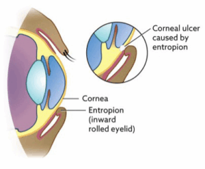
What is entropion?
Entropion is a condition where the eyelids roll inward toward the cornea, which acts as the eye’s windscreen. This inward rolling can cause the eyelashes and hair to rub against the cornea, leading to discomfort, irritation, and potential damage. Common complications include pain, excessive tearing, corneal ulcers, and scarring. In more severe cases, the constant friction can lead to infections or even corneal perforations, which may require urgent medical intervention to prevent vision loss.
What are the signs of entropion?
- Squinting
- Holding the eye shut
- Tearing excessively
- Mucoid discharge
- Keratitis – inflammation of the cornea
- Redness and repeat ‘eye infections’
What types of entropion exist?
- Primary – developmental or hereditary entropion seen at a higher prevalence in certain breeds of dogs and cats.
- Secondary or acquired.
How can acquired (secondary) entropion occur?
This can happen following injury, chronic inflammation, and painful eye disease. Eyelid spasms associated with eye pain (usually ulcerations) results in “spastic entropion” where a vicious cycle associated with squinting and secondary rolling of the eyelid inwards occurs.
In older cats and dogs’ loss of fat behind the eye and age related collagen loss (skin stretching/sagging) can lead to entropion. For example, in older Cocker Spaniels the drooping of the eyelids following a loss of collagen can lead to hair rubbing across the cornea and discomfort. In older cats the loss of fat that sits behind the eyes can lead to sinking of the eye and secondary rolling of the eyelids inwards.
Are certain breeds more likely to have entropion?
Are certain breeds more likely to have entropion? Primary entropion typically develops in dogs up to 2 years of age. If the entropion is identified in young puppies/immature dogs we may opt to perform an interim procedure called ‘eyelid tacking’. This is a temporary correction with sutures to resolve pain and discomfort. This is because entropion may spontaneously resolve when the animal is fully grown and increase our success rate for permanent corrective surgery if required.
How is entropion treated?
The treatment for entropion is surgical correction. There are many techniques used to correct the condition depending on the clinical findings your Ophthalmologist will discuss the technique which is best suited to your pet.
We will often aim to perform corrective surgery once your pet is fully grown however sometimes surgery maybe indicated earlier.
What is the aftercare for entropion?
Both prior to surgery and after surgery, ophthalmic medications such as various antibiotics and artificial tear lubricants may be used to help treat secondary problems that have developed and to protect the cornea, respectively. An Elizabethan Collar will be provided to prevent your pet from rubbing. Usually self absorbable sutures are placed which do not require removal.
What are the possible complications following entropion surgery?
The most common complications involve over or under correction. Under correction may require repeat surgery. Over correction is uncommon when performed by an Ophthalmologist however this still can occur. This may be managed by repeat surgery or medications.
What is the prognosis for entropion?
The prognosis for the surgical correction of entropion is generally good. Occasionally several surgeries may be required to get the best outcome however, most dogs enjoy a pain-free normal life. If the condition is treated later and corneal scarring has occurred, there may be permanent irreversible visual deficits. Your Ophthalmologist will discuss a diagnostic and treatment plan for your dog to help you successfully treat this condition. Overall the success rate for surgery is >95%.
What is ectropion?
Ectropion is an abnormality of the eyelids in which the lower eyelid ‘rolls’ outward or becomes everted. This causes the lower eyelids to appear droopy. Ectropion exposes the pink conjunctival tissue that lines the inner eyelids and cover the white part of the eye. This results in conjunctivitis. The surface of the eye or the cornea may also dry out, resulting in keratitis (corneal inflammation). Corneal damage can also result in corneal scarring, that can impair or obstruct vision. In most cases, both eyes are affected. Ectropion is usually diagnosed in dogs less than one year of age.
What breeds are more likely to have ectropion?
Congenital ectropion is the most commonly seen form. Often in large breed dogs or those with excessive skin. For example, Burnese Mountain dogs, Bulldogs and Spaniels.
Are there other causes of ectropion?
Acquired ectropion can occur in any dog at any age. Acquired ectropion means that a condition other than an inherited trait causes the lower eyelid to ‘sag’ or evert. Some common causes of acquired ectropion include:
- Facial nerve paralysis
- Hypothyroidism
- Scarring secondary to injury
- Chronic inflammation and infection of the tissues surrounding the eyes
- Surgical overcorrection of ectropion
- Neuromuscular disease
What are the clinical signs of ectropion?
- Sagging/rolling outwards of the eyelid (usually the lower eyelid)
- Mucoid discharge
- Redness/inflammation
- Corneal scarring or increased risk of corneal ulcerations
- Tearing and tear staining of the fur
- Repeat ‘eye infections’
- Rubbing/irritation
How is ectropion treated?
The treatment for mild ectropion generally consists of medical therapy, such as lubricating eye drops and ointments to prevent the cornea and conjunctiva from drying out. Ophthalmic antibiotics will be used to combat any corneal ulcers. If the condition is severe, surgical correction can be performed to correct the eyelid position.
What is involved with surgical correction?
Treatment involves eye surgery to restore the normal contour of the eyelid. There are many techniques available, and our Ophthalmologists will discuss which are appropriate for your pet.
How successful is the surgery?
Surgical correction is highly successful, and the prognosis is good. Occasionally under correction can occur and repeat surgery is indicated. The success rate depends on the cause and type of Ectropion.